
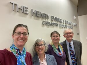
Monday, Apr. 7 & Tuesday, Apr. 8 with W.E. Moerner & Julie Biteen
George and Camilla Smith, generous long-time supporters of the University of Utah, funded the Henry Eyring Lectureship in Chemistry and the Bryant A. Miner Lectureship in Chemistry in 2016.
These endowments memorialize two influential chemists in the Department’s history: Bryant A. Miner, Camilla Smith’s brother, who earned his Ph.D. in chemistry at the U in 1965 and taught countless chemists in his 43 years at Weber State University, and Henry Eyring, Camilla, and Bryant’s uncle, who established the U’s Graduate School and was one of our nation’s most renowned chemists.
These lectures are unique because of Henry Eyring and Bryant Miner’s teacher-student or mentor-mentee relationship. Eyring was the thesis advisor for his nephew’s doctoral degree. The inaugural lectures were held in tandem, featuring two outstanding chemists with the same mentor-mentee relationship. To our knowledge, there is no other program on campus with the same emphasis on this essential academic relationship.
The Smiths’ generosity is an excellent way to recognize Camilla’s uncle and brother and a lasting benefit to future students. These scientists are engaged in challenging yet rewarding fundamental research and teaching, carrying on the legacy of Henry Eyring and the many doctoral graduates of the past 70 years, including Bryant Miner.
The inaugural Henry Eyring and Bryant A. Miner Lectureships were scheduled for March 2017. The Henry Eyring lecture was presented by Nobel Laureate Prof. Robert Grubbs (Caltech), and the Bryant Miner lecturer was presented by former graduate student and professor, Dr. Melanie Sanford (University of Michigan).
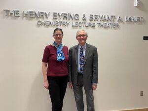
W.E. Moerner
W. E. Moerner, a Professor of Chemistry and Applied Physics at Stanford, has made significant contributions to physical chemistry, biophysics, and super-resolution imaging for cell biology. His work focuses on tracking the motions of proteins, DNA, and RNA in real-time to understand cellular organization and interactions. He has developed 2D and 3D imaging techniques to study viral RNA and proteins, protein structures in bacteria and mammalian cells, and chromatin organization. His previous work involved analyzing the photodynamics of single trapped biomolecules in solution, with applications to photosynthesis and protein interactions.
Moerner earned his B.S. in Physics and Electrical Engineering from Washington University in 1975 and his Ph.D. in Physics from Cornell University in 1982. He worked at IBM before joining UC San Diego as a Distinguished Professor and later the Stanford Chemistry Department in 1997, where he served as Chair from 2011 to 2014.
He won the 2014 Nobel Prize in Chemistry for developing super-resolved fluorescence microscopy, a method that surpasses the optical diffraction limit using single-molecule imaging. His lab focuses on fluorescence microscopy, 3D tracking in cellular environments, and imaging viral infections like SARS-CoV-2. He has received numerous honors, including election to the National Academy of Sciences.
Fun (and Science) with Single-Molecule Optical Microscopy Over the Last 36 Years
Abstract: First observed optically 36 years ago in my laboratory at IBM Research, single molecules have enabled a new field of optical microscopy of the nanoscale. Since ensemble averaging is removed, each single molecule can act as a reporter of not only its position, but also of local information about the nearby environment. Combined with blinking and photoswitching to ensure sparsity (first observed at low temperatures in 1992 and then for single GFP proteins at room temperature in 1997), in the mid-2000’s, super-resolution fluorescence microscopy based on single molecules opened up a frontier in which structures and behavior can be observed in materials and in fixed and live cells with resolutions down to 10-20 nm and below. These methods have been enhanced by PSF engineering to extract 3D position and orientation information, and by deep learning to estimate both molecular variables and structured backgrounds, plus much more. A recent super-resolution study shows fascinating intracellular structures formed by SARS-CoV-2 viral RNA and proteins in infected mammalian cells. Three-dimensional single-molecule tracking in live cells provides time-dependent information about biological regulation and condensed complexes, as well as anomalous diffusion of DNA loci in nuclei.
Another thrust area goes back to isolated single molecules, and explores dynamical behavior in solution in an Anti-Brownian ELectrokinetic (ABEL) trap, with multiparameter extraction of information about complex photodynamics and enzyme kinetics. This trap has recently been extended to non-fluorescent objects with interferometric scattering detection (ISABEL trap), with fluorescence now used as an auxiliary reporter of internal states of carboxysomes.
All of these developments in my laboratory over the past decades have been enabled by an extremely talented cadre of students and postdoctoral researchers, to whom I am very grateful.
Julie Biteen
Julie Biteen is a Collegiate Professor of Chemistry and Biophysics and serves as the Associate Chair of Graduate Education in the Department of Chemistry at the University of Michigan. She has dedicated over 20 years to applying her expertise in optics, imaging, and biophysics to address complex scientific challenges.
Dr. Biteen earned her Ph.D. from the California Institute of Technology and completed postdoctoral research at Stanford University. Her research focuses on bridging the gap between molecular-scale interactions and the traditional limits of light microscopy. Her lab develops single-molecule fluorescence imaging techniques that enable ultrahigh-resolution, real-time imaging, providing insights into biological, physical, and materials science phenomena.
Single-molecule methods, central to her research, have revolutionized multiple disciplines by allowing high-sensitivity measurements, eliminating heterogeneity, and achieving nanometer-scale resolution. These techniques are particularly impactful in microbial cell biology and metal nanoparticle plasmonics.
Beyond scientific inquiry, Dr. Biteen is dedicated to defining what it means to be a scientist in the 21st century. The Biteen Lab serves as a safe, inclusive space where individuals from diverse backgrounds can thrive both as scientists and community members. Dr. Biteen is committed to fostering a welcoming environment, ensuring that marginalized groups are empowered to succeed in science.
Determining the nature of molecular interactions and biomolecular condensates in microbes
Abstract: Single-molecule microscopy accesses nanometer-scale information with a benchtop microscope, providing a platform to super-resolve fluorescence emission, position, and dynamics, even in living cells. The Biteen Lab is developing new single-molecule methods to answer fundamental, unanswered questions in microbiology with applications including elucidating cell regulation and misregulation, understanding epigenetic inheritance, and visualizing nutrient utilization in the microbiome. These direct, quantitative, and high-resolution approaches have consequences in understanding subcellular biochemistry and biophysics. I will focus on our recently developed approaches to quantifying how cellular components interact and organize in microbiology. On one hand, we are evaluating the millisecond- and nanometer-scale dynamics of specific partners, and, as an example, I will present how HP1 proteins specifically and selectively associate with heterochromatin to silence gene expression in fission yeast. On the other hand, we are developing a generalizable, accessible, and rigorous framework to probe the nature of biomolecular condensates on the sub-micron scale in bacterial cells, and I will show how we probed the formation, reversibility, protein dynamics, and material state of biomolecular condensates in Escherichia coli to achieve a general model of bacterial cell organization.
History of Eyring-Miner Lecture Speakers at the University of Utah
2022-2023
Graham Cooks, Purdue University
Some Discoveries in Mass Spectroscopy
Livia Eberlin, Baylor College of Medicine
Guiding Medical Decisions with Mass Spectrometry Technologies
2019-2020
Suzanne A. Blum, UC Irvine
Mechanistic Chemistry at the Single-Molecule Level
Robert G. Bergman, Berkeley
A Nostalgia Trip through Organometallic Chemistry of the Late 20th Century
2018-2019
Squire J. Booker, Penn State
Moving beyond Methionine Synthase: New Insights Into Cobalamin-Dependent Methyltransferase Reactions
JoAnne Stubbe, MIT
Radicals: Your life is in their hands
2017-2018
Christy Haynes, University of Minnesota
Design and Redesign of Sustainable Engineered Nanomaterials
George Schatz, Northwestern
2016-2017
The Inaugural Speaker of the Lecture
Robert Grubbs, California Institute of Technology
Design and Applications of Selective Olefin Metathesis Catalysts
Melanie Sanford, University of Michigan
Development and Applications of New Fluorination Reactions